
Cochlea – overview, labeling arrows
Image
Unlabeled graphic:
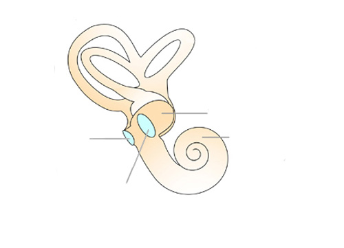
The structure of the cochlea is shown. Labeling arrows at: oval window, round window, vestibule and cochlea.
English, Spanish (CREA), German
2018-04-22

This medium is made available under a CC BY-SA 4.0 international license.
What does this mean?
How to reference this medium
The cochlea consists of a coiled canal which appears in three compartments in the section. The part leading upwards is called the scala vestibuli and begins at the oval window.
The part leading downwards is called the scala tympani.
Between scala vestibuli and scala tympani there is a membranous tube which is also filled with fluid. This is where the actual hearing organ, the Organ of Corti is located.
Information and ideas:
Can be used in a worksheet, for work together on the digital projector, or as an overhead transparency.
Further information regarding this graphic labeling is available as information sheet on the media portal of the Siemens Stiftung.
Relevant for teaching:
Structure and functions of a sensory organ
Reception of stimuli and transmission of information
Illustration
Biology
Grade 5 to 6; Grade 7 to 9; Grade 10 to 13
Middle/high school; Vocational training
Anatomy (human); Ear; Ear (inner ear); Medical illustration
Siemens Stiftung Media Portal
MediaHouse GmbH
© Siemens Stiftung 2018