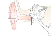
Outer ear section – labeling arrows
Image
Unlabeled graphic:
The graphic is a section view of the entire ear showing all parts belonging to the outer ear. These parts are highlighted in color.
Type of media:
Image (81.9 kByte)
Last update:
2018-02-01
License:

This medium is made available under a CC BY-SA 4.0 international license.
What does this mean?
How to reference this medium

This medium is made available under a CC BY-SA 4.0 international license.
What does this mean?
How to reference this medium
Media package:
Description:
The outer ear consists of the pinna and the ear canal. The ear canal ends at the eardrum.
In the membranous wall of the ear canal there are glands which produce cerumen (earwax). At the edge of the ear canal there are some small hairs, hair follicles, which serve as protection against foreign bodies.
Information and ideas:
Helpful to distinguish outer, middle and inner ear.
Can be used, for example in a worksheet, for work together in class with the digital projector, as overhead transparency.
Relevant for teaching:
The human body
Structure and function of a sensory organ
Learning resource type:
Illustration
Subjects:
Biology
Grade levels:
Grade 5 to 6; Grade 7 to 9; Grade 10 to 13
School types:
Middle/high school; Vocational training
Keywords:
Anatomy (human); Ear; Ear (outer ear); Medical illustration
Bibliography:
Siemens Stiftung Media Portal
Author:
MediaHouse GmbH
Rights holder:
© Siemens Stiftung 2016


