
Middle ear section – labeling arrows
Bild
Unlabeled graphic:
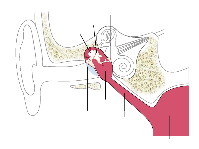
Section view of human ear with the individual parts of the middle ear – with arrows for detailed labeling.
Englisch, Deutsch, Spanisch (CREA)
01.02.2018

Dieses Medium steht unter einer CC BY-SA 4.0 international Lizenz.
Was bedeutet das?
So verweisen Sie auf das Medium
The middle ear is formed by an air-filled cavity lined with mucous membrane and consists mainly of the tympanic cavity and the Eustachian tube.
The tympanic cavity contains the ossicles “malleus", “incus" and “stapes".
These are joined together loosely and can move so that, with their help, vibrations from the eardrum can be picked up and transmitted to the inner ear.
Information and ideas:
Can be used in worksheet, worked on together via digital projector, as an overhead transparency.
Relevant for teaching:
The human body
Structure and function of a sensory organ
Middle ear section (Bild)
Illustration
Biology
Grade 5 to 6; Grade 7 to 9; Grade 10 to 13
Middle/high school; Vocational training
Anatomy (human); Ear; Ear (middle ear); Medical illustration
Siemens Stiftung Media Portal
MediaHouse GmbH
© Siemens Stiftung 2016