
Cochlea – transparent uncoiled
Bild
Graphic:
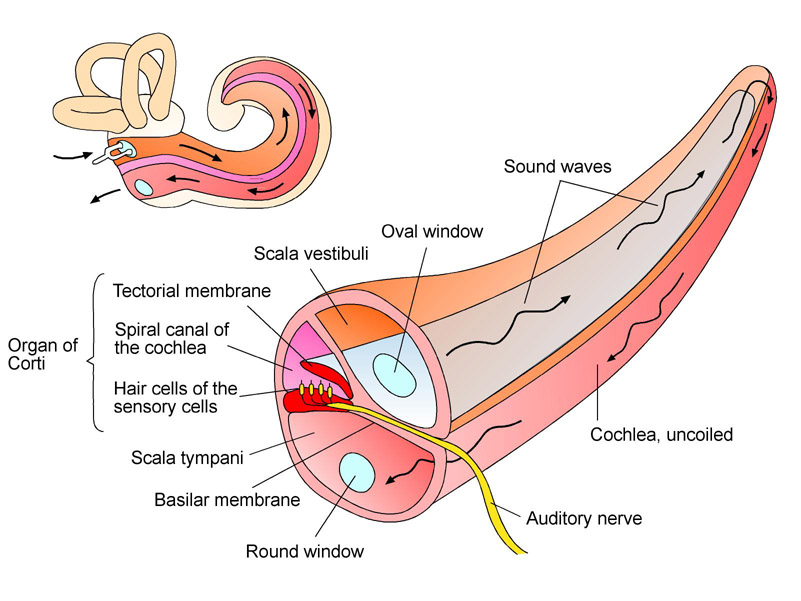
Spatially transparent cross-section of the uncoiled cochlea with scala vestibuli, scala tympani and spiral canal of the cochlea.
Englisch, Spanisch (CREA), Deutsch
27.07.2018

Dieses Medium steht unter einer CC BY-SA 4.0 international Lizenz.
Was bedeutet das?
So verweisen Sie auf das Medium
The flow direction of sound as a traveling wave is sketched in. The position of the organ of Corti as sound recipient is shown as well.
Furthermore it is clearly illustrated that the scala tympani and the scala vestibuli are one single fluid cavity.
Information and ideas:
This graphic helps to make clear that the whole cochlea is a fluid canal and that it is there where the vibrations of sensory hair cells are converted into nerve impulses.
Can be used on worksheets or as overhead transparency.
Relevant for teaching:
Structure and functions of a sense organ
Reception of stimuli and transmission of information
Functions of senses
Illustration
Biology
Grade 5 to 6; Grade 7 to 9; Grade 10 to 13
Middle/high school; Vocational training
Anatomy (human); Ear; Sound; Ear (inner ear); Medical illustration; Sound transduction
Siemens Stiftung Media Portal
MediaHouse GmbH
© Siemens Stiftung 2018
